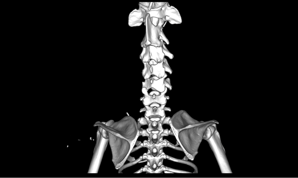
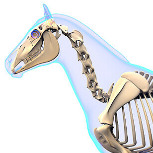

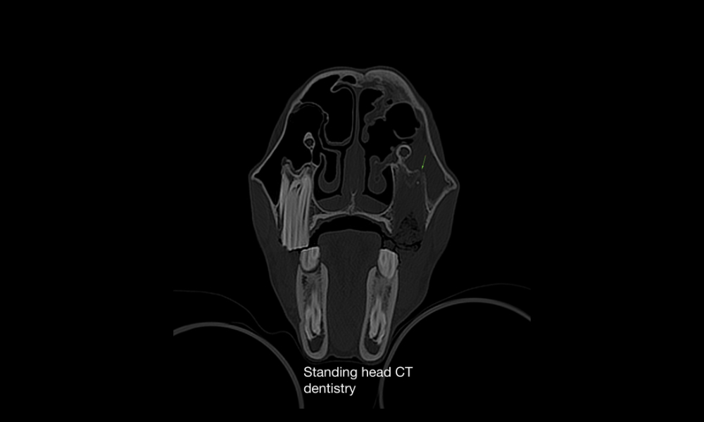
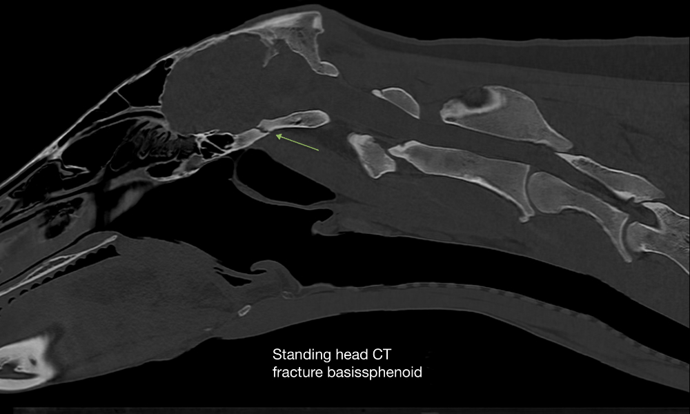
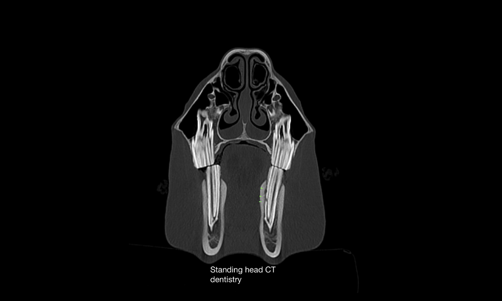
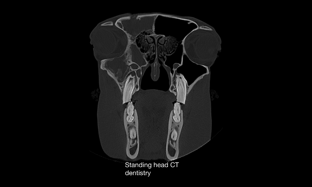
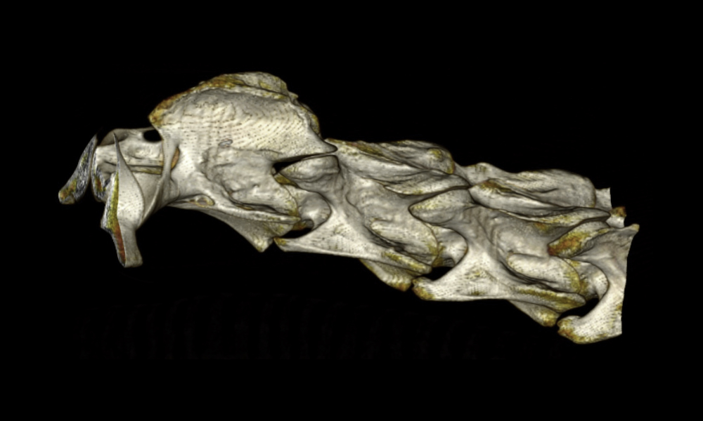
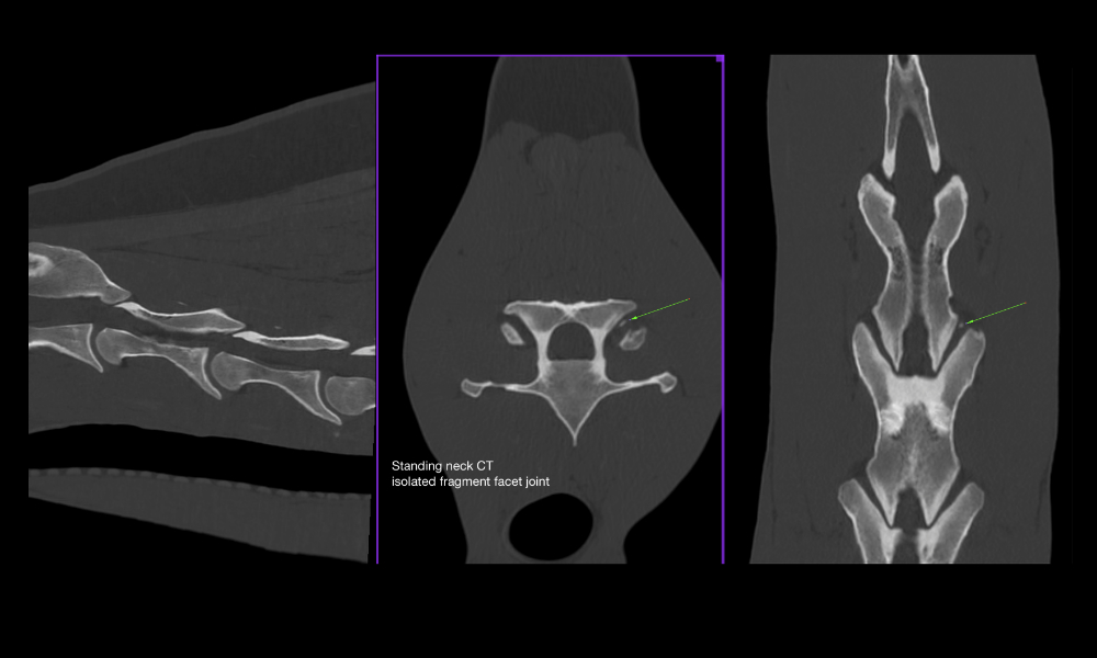
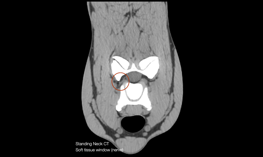
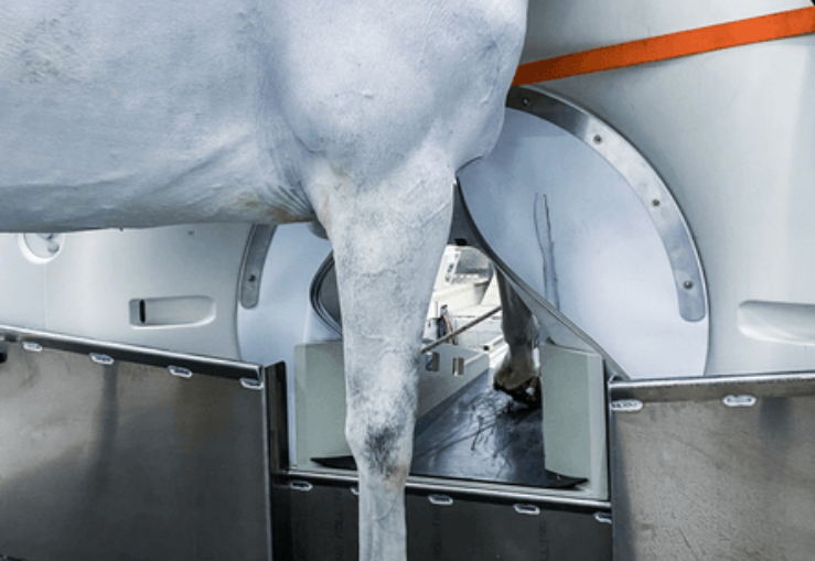
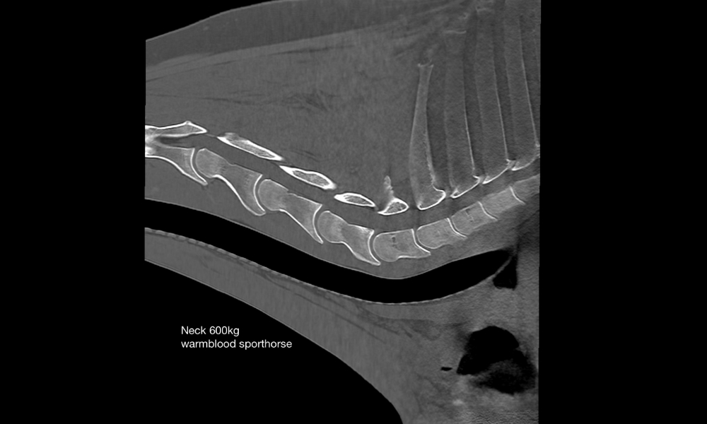
Head / neck of the standing horse
- Simple, safe, and efficient patient positioning
- Examinations can be completed in minutes
- 0,5mm thin slices allow visualization of even the thinnest bone lamellae, periodontal supporting ligaments and intervertebral nerves
- The neck can be depicted up to C5/6 in a standing horse
- The modified front cover shortens the distance to the isocenter
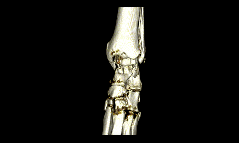
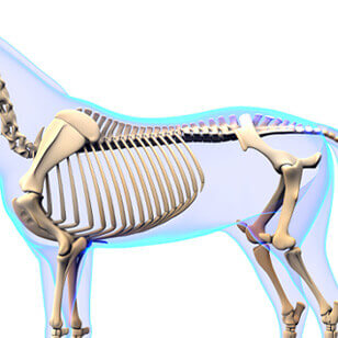
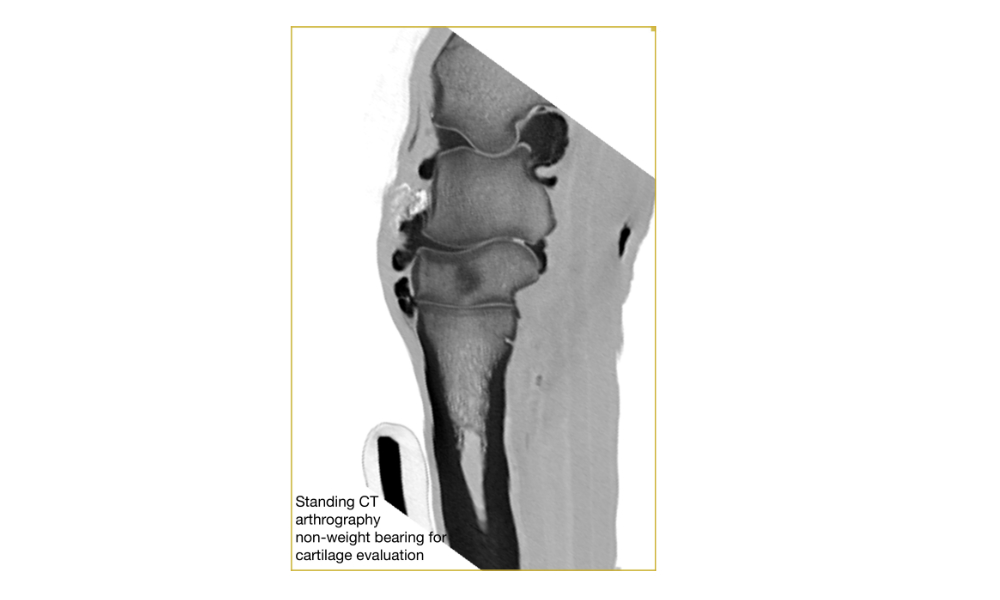
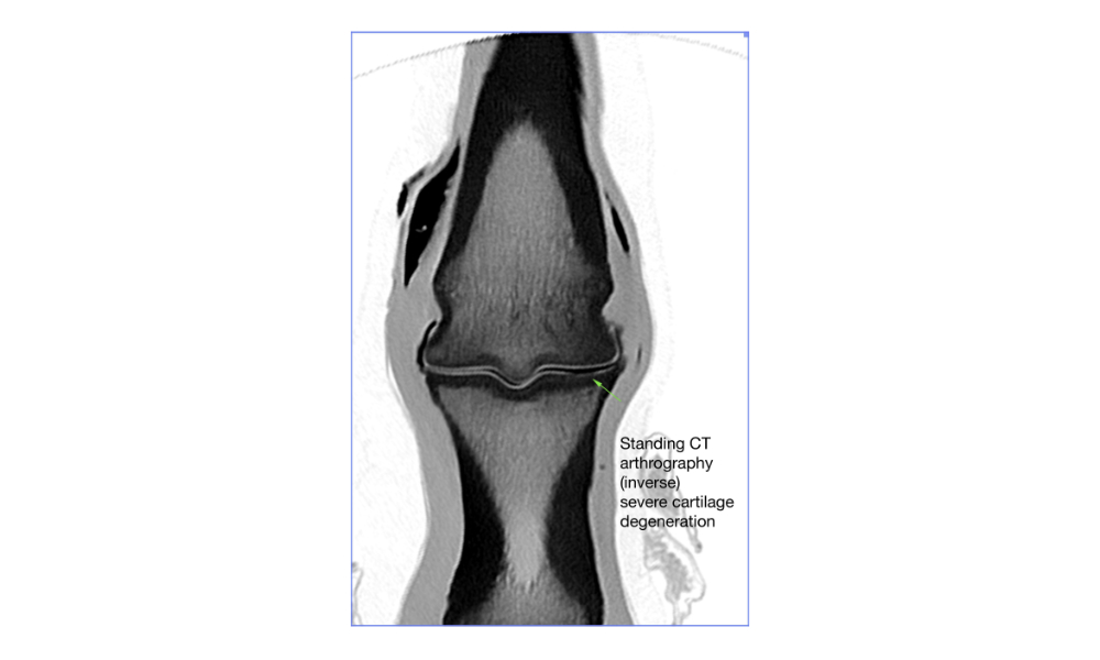
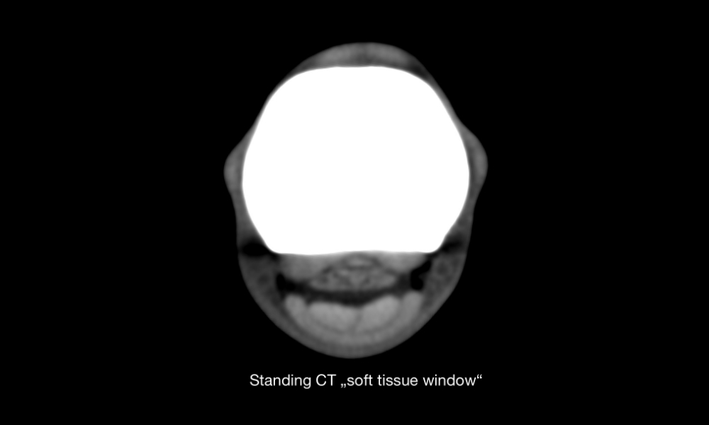
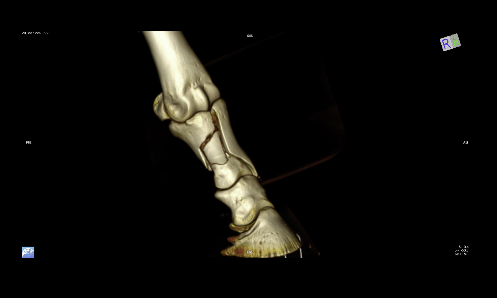
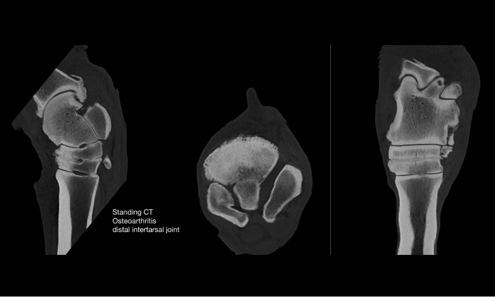
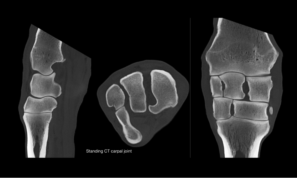
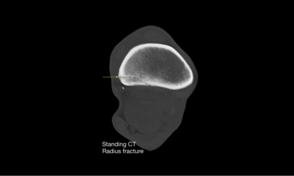
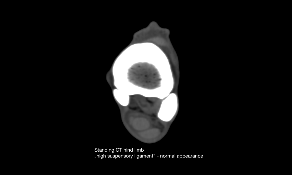
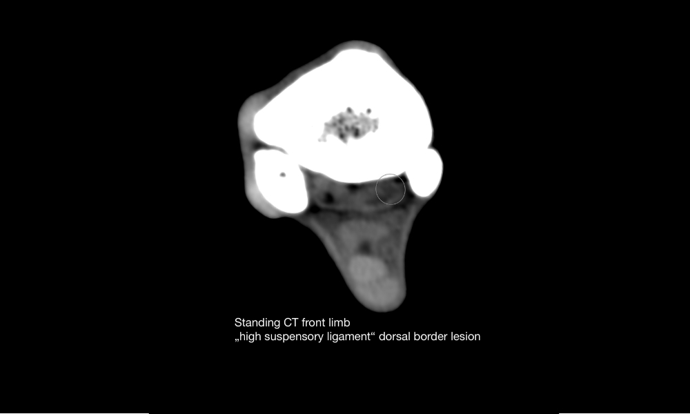
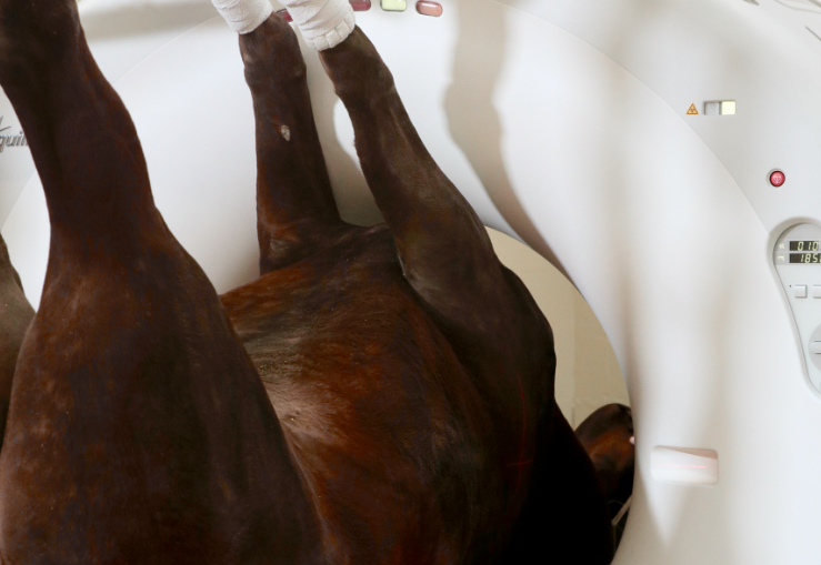
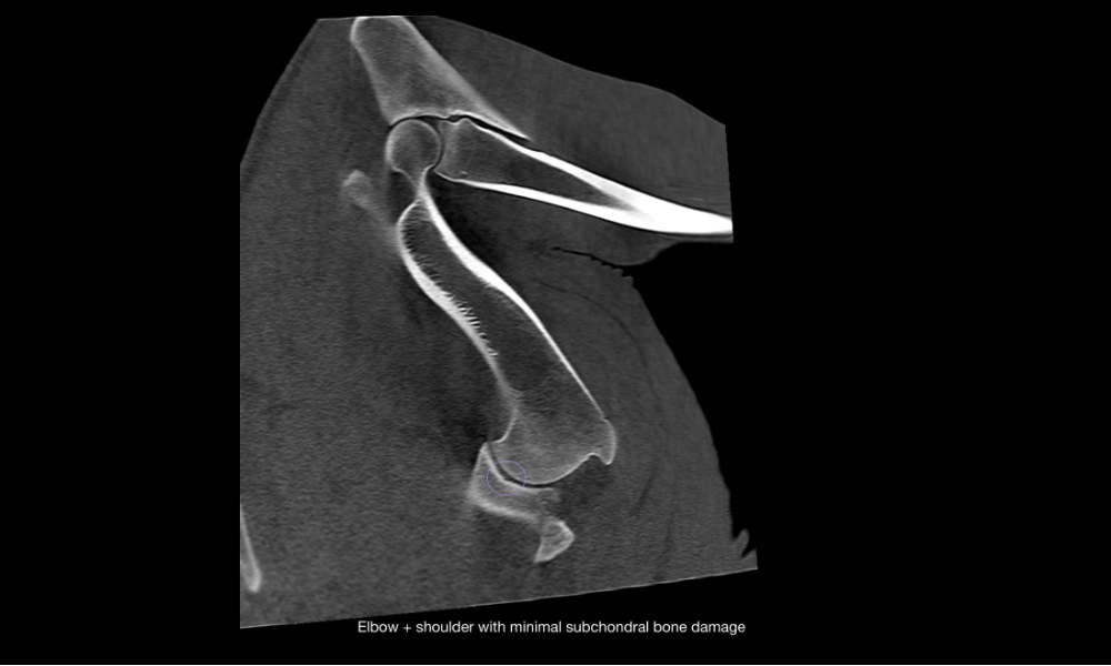
Distal limb CT of the standing horse
- Examinations up to the radius or the tibia
- Incredibly short examination times as quick as 0.28 sec/16 cm (system dependent)
- Superior soft tissue detection compared to low-field MRI
- Positioning of the horse is similar to farrier services, providing familiarity and comfort
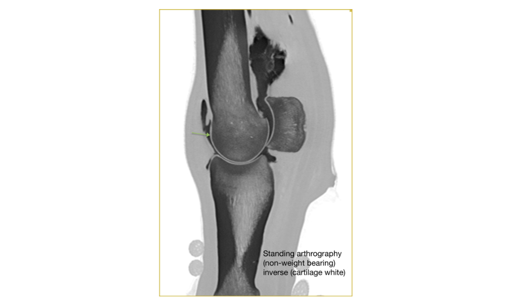
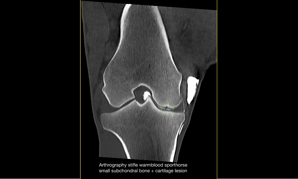
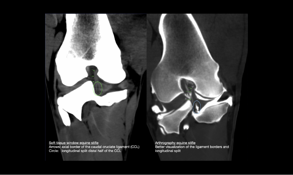
- Standing, yet non weight bearing examinations allow CT arthrography without general anesthesia and reduce artifacts from load bearing
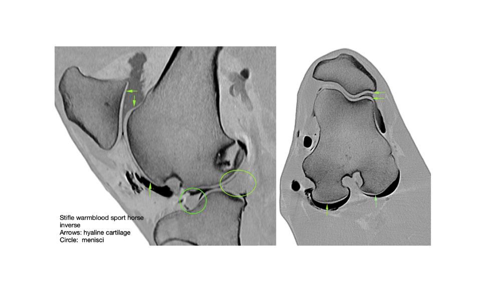
- The 0.5mm slice size combined with the latest algorithms and over 80 reconstruction filters allow imaging of the smallest lesions in ligaments, tendons and hyaline cartilage

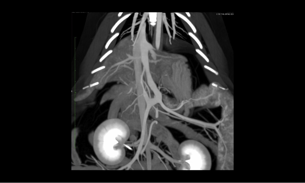
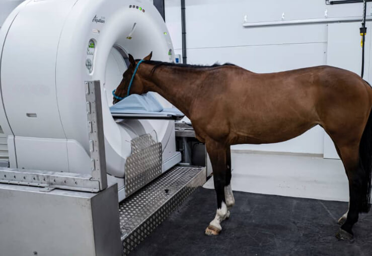
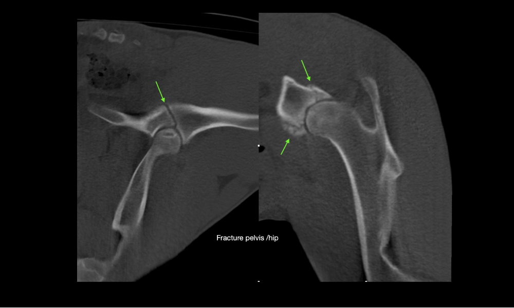
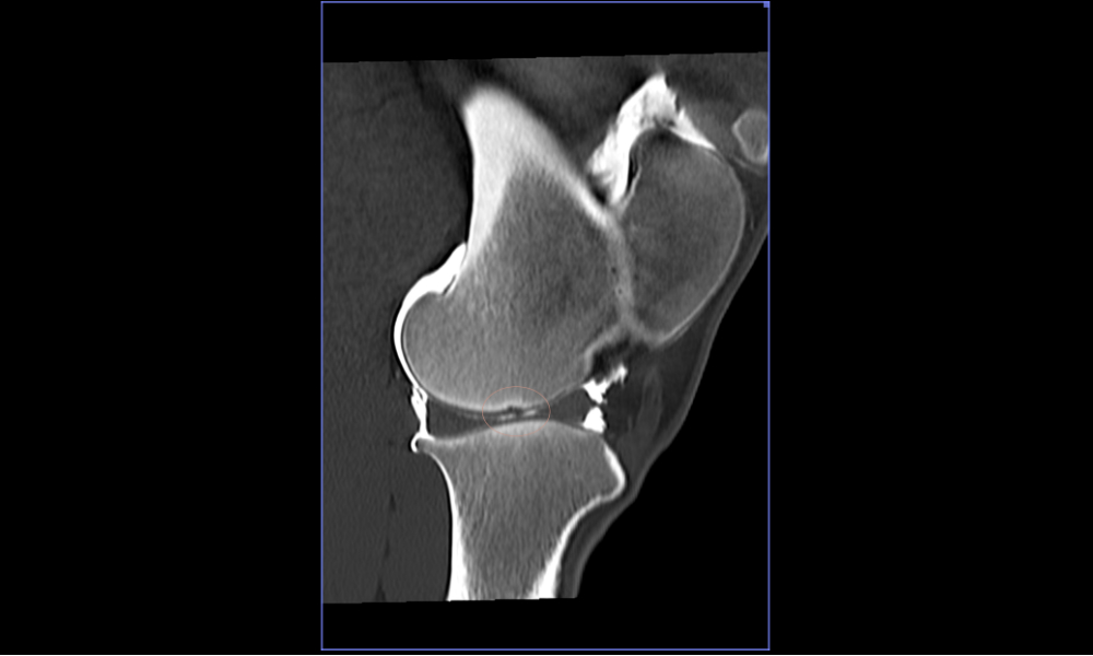
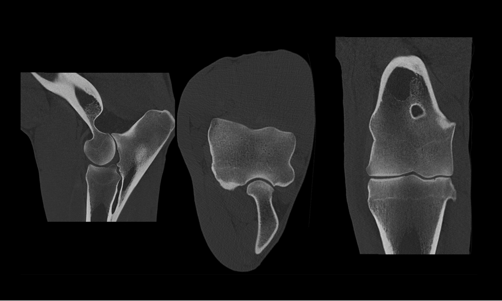
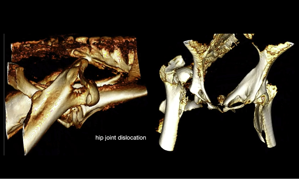
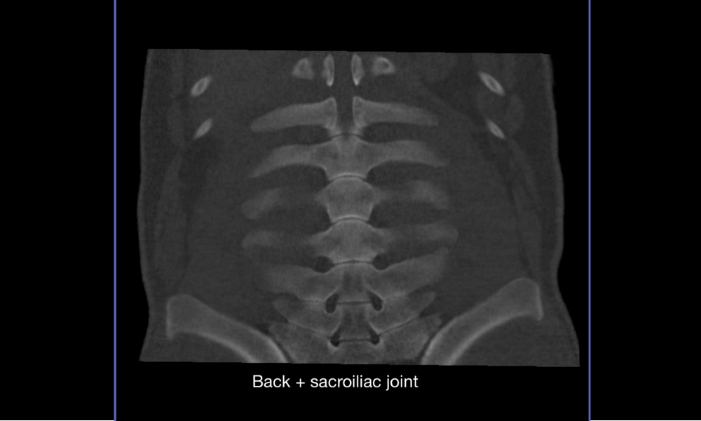
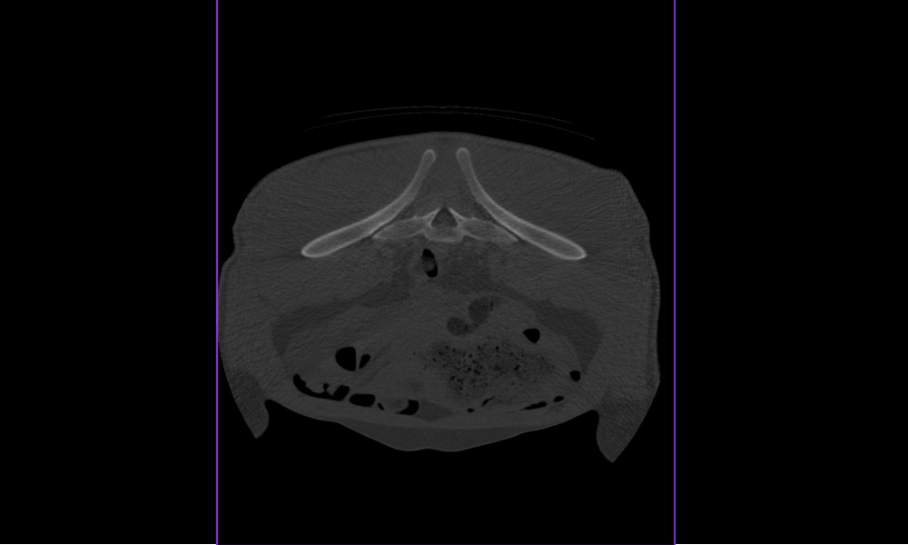
Pelvis, back, stifles, etc. in the anesthetized horse
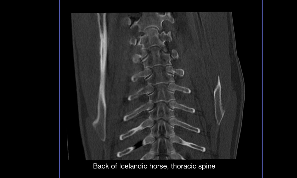
- Our large bore system offers the largest gantry opening (90cm) available in medical practice, permitting examinations of the equine thoracic spinal column, the back and hip joints
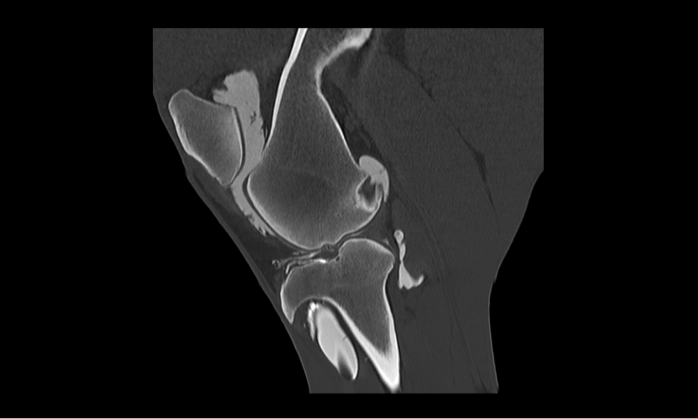
- Simplified positioning of the horse without an external table significantly shortens the duration of anesthesia (e.g., CT of both stifle joints in under 15 minutes)
- The gantry moves over the horse, no need to pull the horse into the gantry for positioning
- Large field of view (up to 90 cm), high generator output and software with the latest interactive algorithms make examining areas with high X-ray density, such as a large body mass, possible (e.g. parallel examination of both knees)
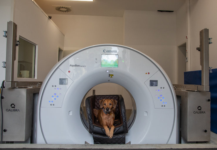
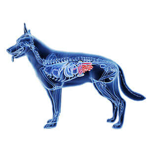
Small animals
- The unique configuration of the Qalibra CT system allows the gantry to be lowered to the ground level, permitting scanning of the patient without sedation
- Since the couch is immobile, there is no need for modified anesthesia equipment.
- Advanced imaging solutions (e.g., dose modulation and bolus tracking) are available and with some units, volume scans of up to 16cm are possible in less than 1/3 of a second
- Lead shielding enables performing CT scans in awake patients, similar to performing an X-ray