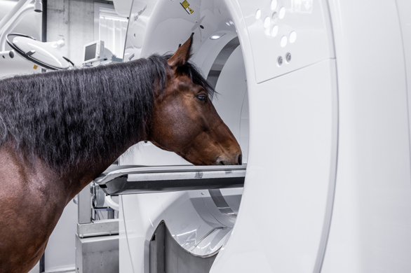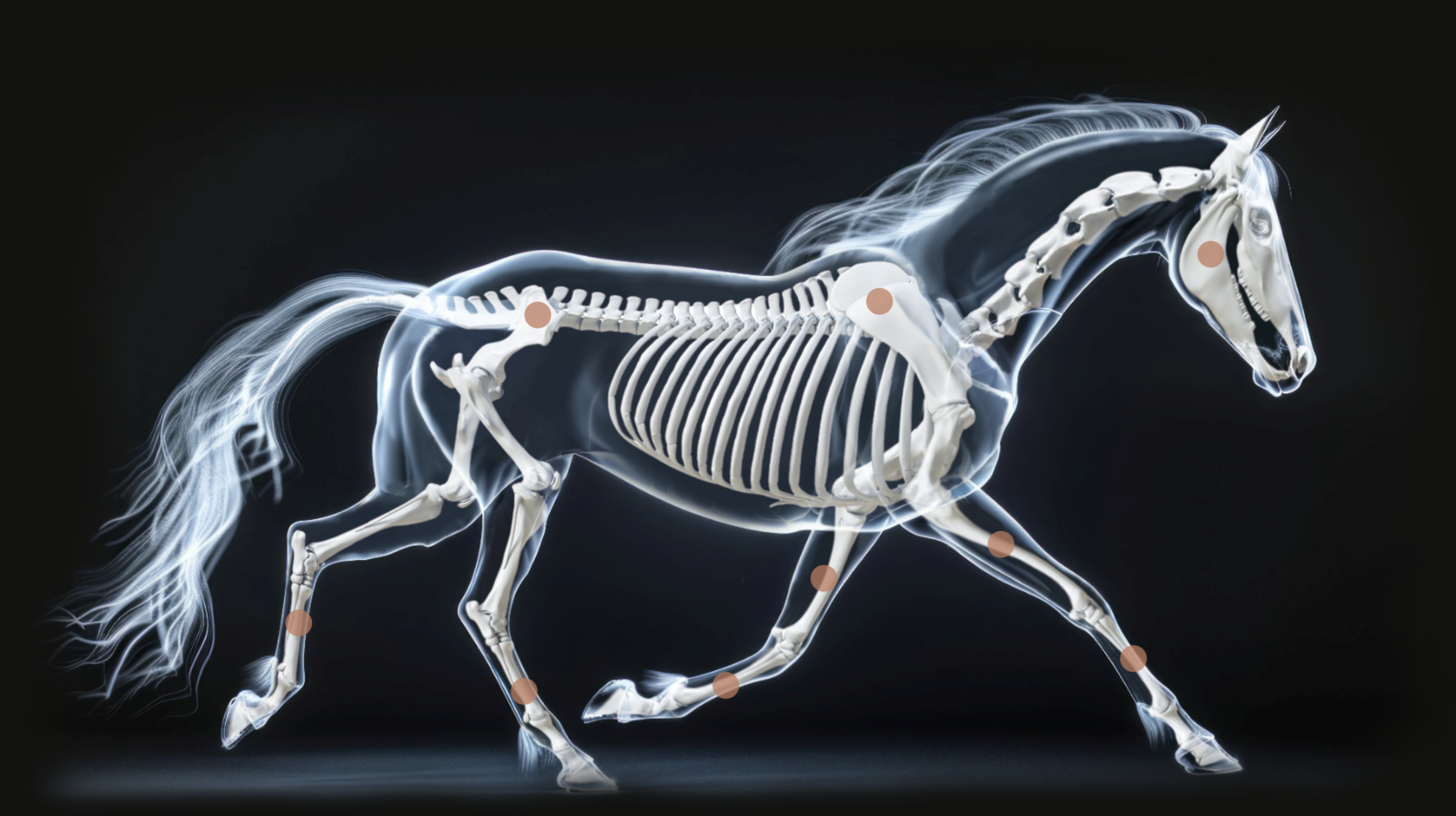
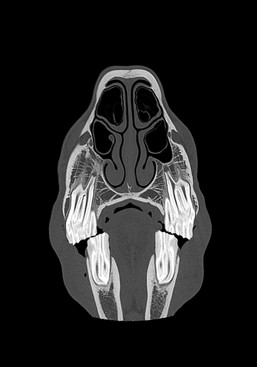
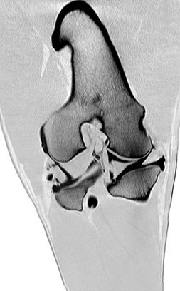
Head / neck of the standing horse
- Simple, safe, and efficient patient positioning
- Examinations can be completed in minutes
- 0,5mm thin slices allow visualization of even the thinnest bone lamellae, periodontal supporting ligaments and intervertebral nerves
- The neck can be depicted up to C5/6 in a standing horse
- The modified front cover shortens the distance to the isocenter

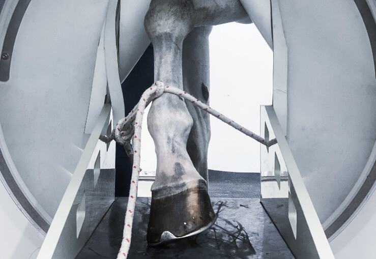
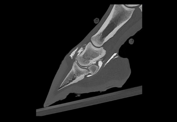
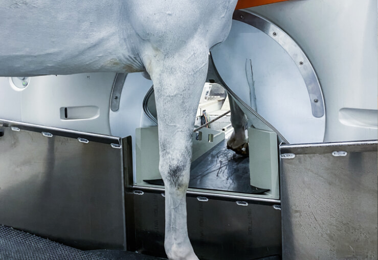
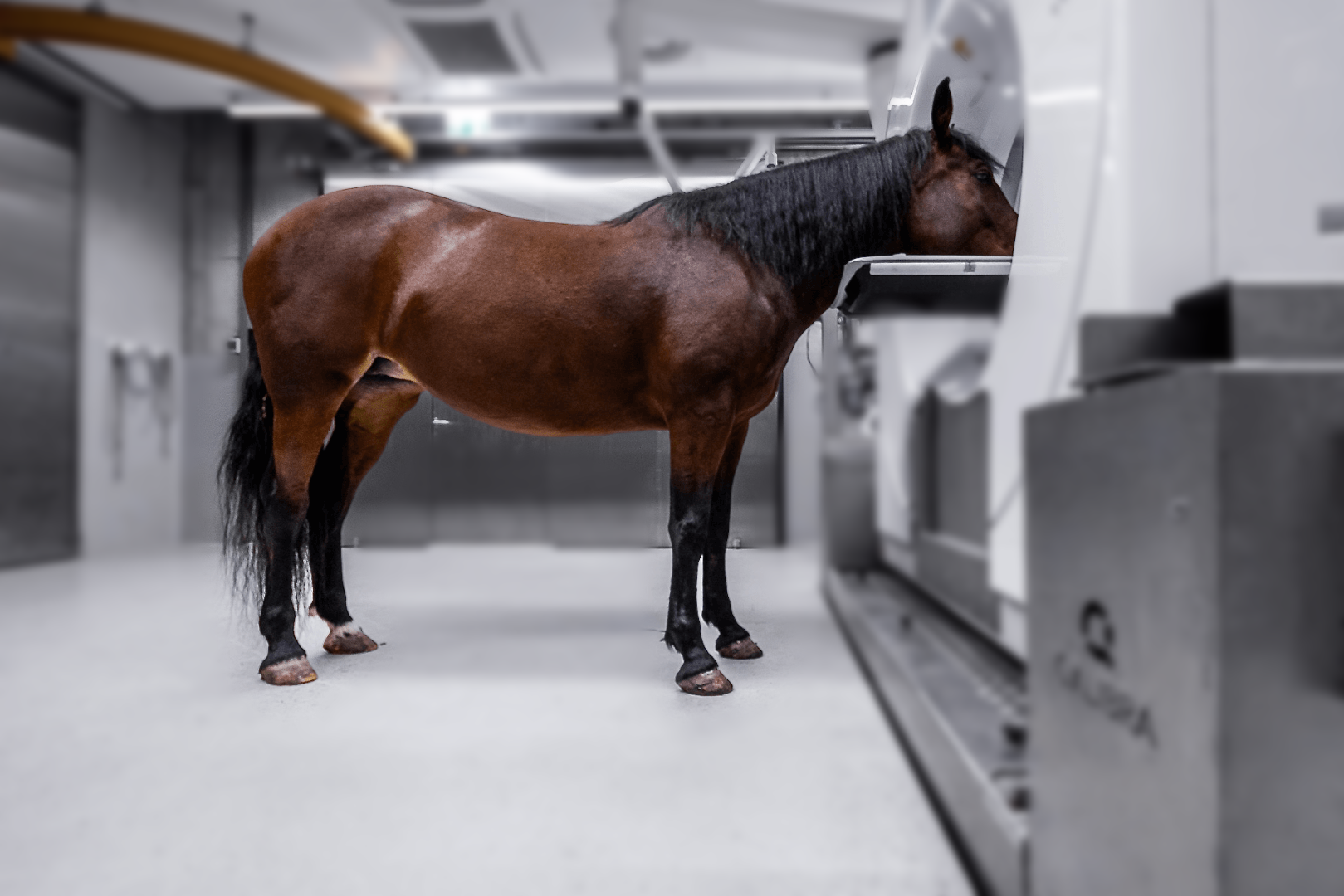
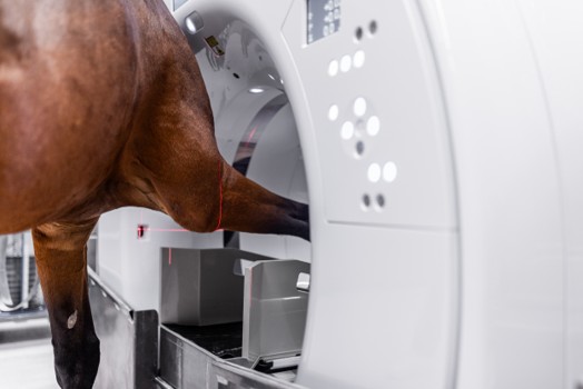
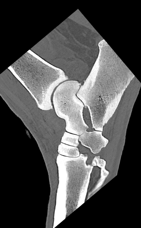
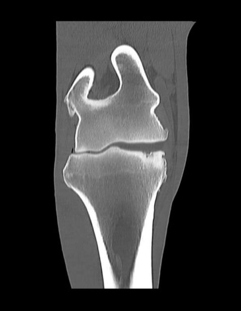
Distal limb CT of the standing horse
- Examinations up to the radius or the tibia
- Superior soft tissue detection compared to low-field MRI
- Positioning of the horse is similar to farrier services, providing familiarity and comfort
Distal limb CT of the standing horse
- Examinations up to the radius or the tibia
- Superior soft tissue detection compared to low-field MRI
- Positioning of the horse is similar to farrier services, providing familiarity and comfort
Distal limb CT of the standing horse
- Examinations up to the radius or the tibia
- Superior soft tissue detection compared to low-field MRI
- Positioning of the horse is similar to farrier services, providing familiarity and comfort
Distal limb CT of the standing horse
- Examinations up to the radius or the tibia
- Superior soft tissue detection compared to low-field MRI
- Positioning of the horse is similar to farrier services, providing familiarity and comfort
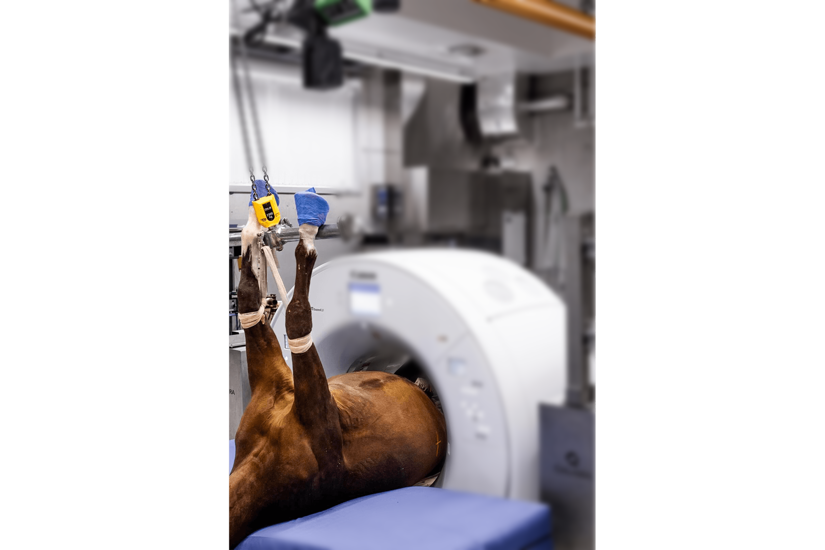
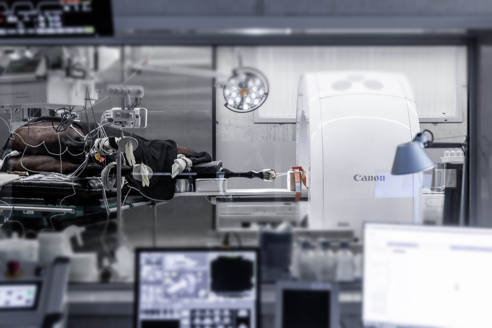
Pelvis, back, stifles, etc. in the anesthetized horse
- The gantry moves over the horse, no need to pull the horse into the gantry for positioning
- Simplified positioning of the horse without an external table significantly shortens the duration of anesthesia (e.g., CT of both stifle joints in under 15 minutes)
- Simple, safe, and efficient patient positioning
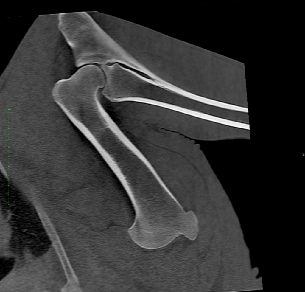
Elbow and forearm CT of the standing horse
- Examinations up to and including the elbow and proximal radius/ulna
- Excellent visualization of bone and soft tissue structures in a non-weight-bearing position
- Positioning is simple and familiar, similar to routine limb handling
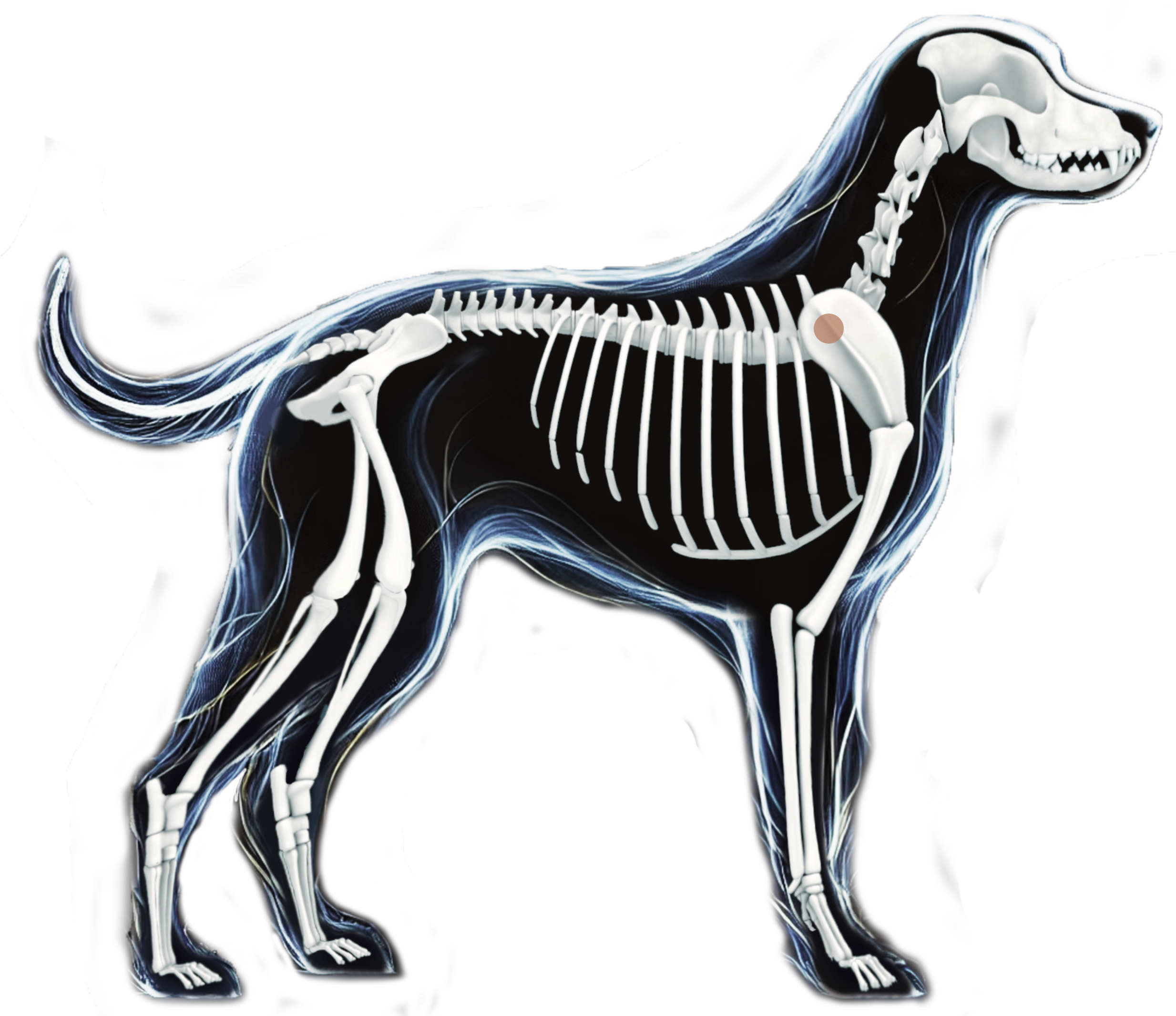
Shoulder CT of the horse
- Detailed imaging of the scapulohumeral joint and surrounding soft tissues
- General anesthesia required due to joint depth and anatomical positioning
- Ideal for evaluating cartilage lesions, joint pathology, and scapular injuries
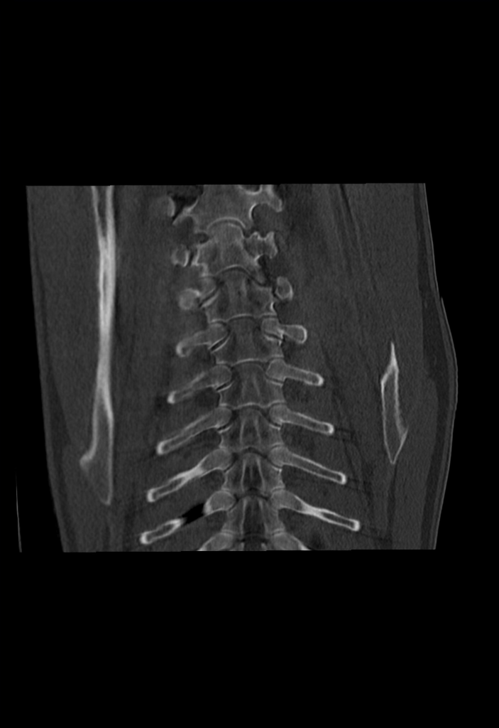
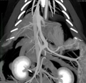
Thoracic region CT of the horse
- High-resolution imaging of lungs, ribs, and cranial thoracic spine
- General anesthesia ensures optimal image quality and eliminates motion artifacts
- Indicated for detecting pulmonary lesions, pleural effusion, or rib fractures
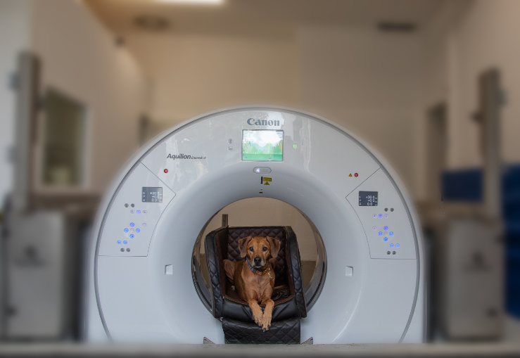
Small animals
- Since the couch is immobile, there is no need for modified anesthesia equipment.
- Lead shielding enables performing CT scans in awake patients, similar to performing an X-ray
- The unique configuration of the Qalibra CT system allows the gantry to be lowered to the ground level, permitting scanning of the patient without sedation